Reporter: Claudia Villalobos | Photographer: Enrique Lair
IPN researcher develops highly accurate 3D-printed breast models to support medical diagnosis.
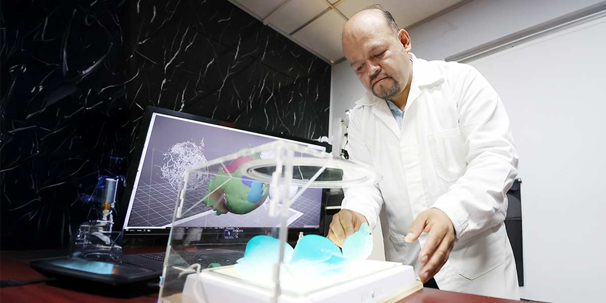
Tangible models, such as those created by researcher Juan Alfonso Beltrán Fernández, enable specialists to make more informed clinical decisions.
From computed tomography (CT) scans, Juan Alfonso Beltrán Fernández, a researcher at the Instituto Politécnico Nacional (IPN), generates digital breast models that are then 3D-printed with remarkable accuracy. These replicas of internal breast structures provide physicians with a complementary tool to improve early breast cancer detection and diagnosis.
The project, still at the research stage, introduces a novel technique: experimental photopolymer resin is exposed to a polarizing lens, which enhances color contrast in areas where tumor tissue is present. This innovation can aid in early detection, improve surgical planning, strengthen medical training, and help raise patient awareness.
Dr. Beltrán Fernández, a biomechanical engineer at the Escuela Superior de Ingeniería Mecánica y Eléctrica (ESIME), Zacatenco campus, emphasized that tangible models are invaluable to physicians, as they allow for more precise and timely decision-making in patient care.
Because this approach represents a pioneering diagnostic tool, Dr. Beltrán Fernández presented the research at the 18th International Conference on Advanced Computational Engineering and Experimentation (ACEX 2025), held in June in Naples, Italy.
A Level II member of Mexico’s National Researchers System (SNII), Dr. Beltrán Fernández, explained that the models are generated by processing CT scans, MRIs, mammograms, and even ultrasound images through ScanIP software. This program produces a 3D-printable stereolithographic (STL) file.
Using an additional visualization tool, Meshmixer, researchers can dynamically isolate, manipulate, and study each breast structure—from the skin and nipple to ducts, vessels, lobules, tissues, and lymph nodes. Within these layers, atypical tissues often associated with tumors can be identified.
Unlike conventional scans, which rely on grayscale interpretation, this software enables each anatomical structure to be visualized independently.
The unique properties of the photopolymer resin make tumor detection possible in the printed models.
Once the digital model is complete, it is 3D-printed in photopolymer resin. “The internal physiology of the breast is reproduced at approximately 80 to 100 percent of its actual size. The process takes around eight to nine hours,” explained Dr. Beltrán Fernández.
He noted that the photopolymer resins used in these prototypes have highly sensitive optical properties. When placed in a custom-designed portable polarizer and exposed to white light, the models can be examined in detail to detect potential tumor formations, which appear as darker regions.
When viewed under the polarizer, small spots with green or iridescent patterns become visible. These indications may guide physicians in deciding whether or not a biopsy is necessary.
Looking ahead, the project could be applied in hospitals across the country, with the possibility of developing more flexible materials to assist in surgical planning.

INSTITUTO POLITÉCNICO NACIONAL
D.R. Instituto Politécnico Nacional (IPN). Av. Luis Enrique Erro S/N, Unidad Profesional Adolfo López Mateos, Zacatenco, Alcaldía Gustavo A. Madero, C.P. 07738, Ciudad de México. Conmutador: 55 57 29 60 00 / 55 57 29 63 00.
Esta página es una obra intelectual protegida por la Ley Federal del Derecho de Autor, puede ser reproducida con fines no lucrativos, siempre y cuando no se mutile, se cite la fuente completa y su dirección electrónica; su uso para otros fines, requiere autorización previa y por escrito de la Dirección General del Instituto.
